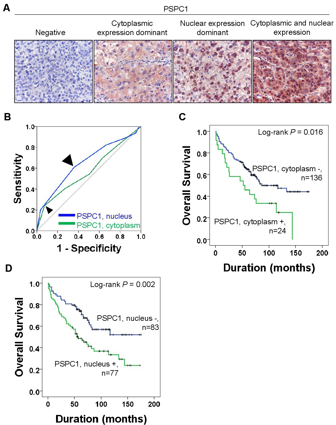
Fig. 6. Immunohistochemical detection of PSPC1 and survival analysis in 160 hepatocellular carcinoma samples. A. Representative pictures of immunohistochemical staining of PSPC1 regarding cytoplasmatic and nuclear staining of tumor cells. B. Statistical analysis to determine cut-off points for cytoplasmic and nuclear expression of PSPC1 with receiver operator characteristic curve analysis. The points with the highest area under the curve (AUC) were cut-off points for cytoplasmic and nuclear PSPC1 expression. C. Kaplan-Meier survival analysis according to cytoplasmic PSPC1 expression and nuclear PSPC1 expression for the overall survival and relapse-free survival.